EN
  • International

  • English Version EN
Pain in the ankle joint

Pain in the ankle joint

The ankle joint

We all take more than 200 million steps throughout our lives. Each of those steps is no mean feat. What looks so simple is actually a complex sequence of movements with perfect timing and constant synchronisation. The ankle, as a pivot point, allows a wide range of movements and copes with all kinds of demands.

The ankle joint is always in motion and also subject to the constant impact of strong forces. That makes it all the more important to treat injuries and any subsequent ligament instabilities properly at an early stage in order to prevent serious consequential damage.


Anatomy of the ankle joint  

The ankle joint is perfectly designed to fulfil its various functions and is characterised by an ideal interplay between the bones, muscles, ligaments and tendons.  

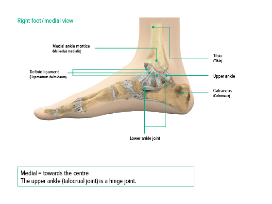
The ankle joint consists of two partial joints: the upper ankle joint and the lower ankle joint. These partial joints supplement each other’s functionalities, forming one unit. This interaction enables lifting and lowering as well as rotating in the direction of the inner and outer edge of the foot (supination and pronation).  


Ankle, medial view

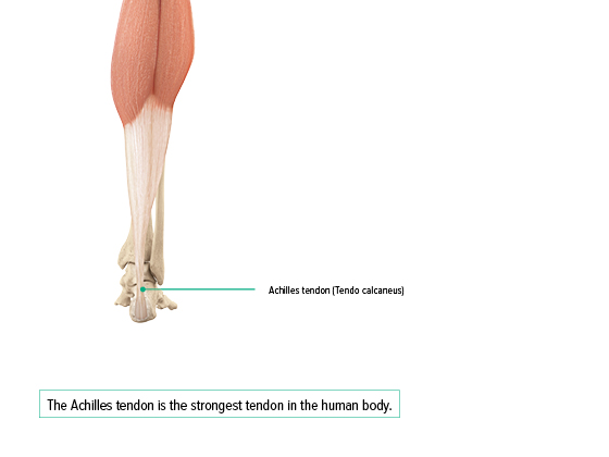
As the body’s strongest tendon, the Achilles tendon connects the flexor muscles of the thigh to the foot bones, thus enabling the foot to flex. It is approximately 5 cm wide and 20 to 25 cm long.

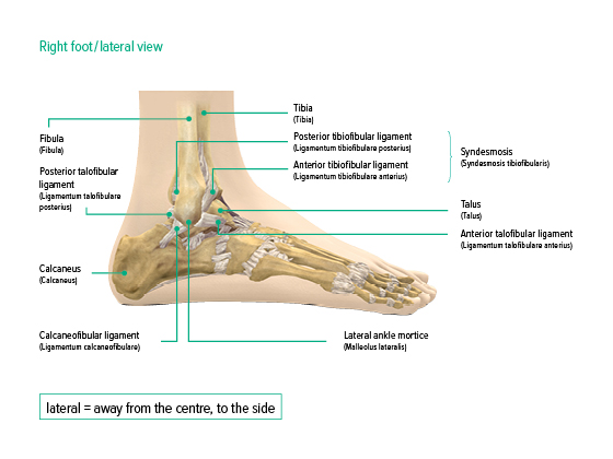
Ankle joint, lateral view

The ankle joint is stabilised by a strong system of ligaments. The elasticity of ligaments is limited to just 3% of the original length. Three individual ligaments secure the ankle joint from the exterior (posterior, central and anterior lateral collateral ligament). The inside is strengthened by a fan-shaped medial collateral ligament complex (deltoid ligament).  

There is also an anterior and posterior tibiofibular ligament. These elastic connective tissue structures connect the tibia and the fibula to the ankle where they play the part of an artificial joint.  

The talar bone (lat. talus) plays a central role in the transmission of force from the lower leg to the foot. If the bones in the foot joint can be moved towards the toes while the lower leg stays in position, this is referred to as talar shift.  

Achilles tendon

Causes of pain in the ankle joint  

Pain in the ankle joint usually occurs as a result of an acute injury. However, other causes can also lead to pain in the ankle joint, such as misalignments, irritated tendons, inflammation, excess strain, inherited factors like congenital weak ligaments as well as conditions caused by wear, e.g. osteoarthritis.


Diagnosing an ankle injury

The basis of a diagnosis is a clinical examination. A tentative diagnosis includes taking a medical history, looking at and palpating the affected area as well as a mobility and stability test. Talar shift, for example, is detected in what is known as the drawer test. The tentative diagnosis is usually verified afterwards using technical imaging methods, such as X-ray, ultrasound scans, MRI and CT.

Man at a medical consultation

Localisation of pain  

The area where pain is felt can be an indication of the type of injury or condition.


  • Pain in the outer ankle indicates an injury to the lateral collateral ligament, a fracture of the outer ankle or a peroneal tendon condition.
  • Pain in the inner ankle can be caused by an injury to the medial collateral ligament, a fracture of the inner ankle, Osteochondrosis dissecans, tarsal tunnel syndrome or lesions of the tibialis posterior tendon.
  • In cases of damage to the lower ankle joint, pain is usually projected to the outer edge of the foot.
  • Pain in the upper ankle joint that mostly occurs during movement indicates an impingement syndrome.
  • If pain is more wide-spread and cannot be pinned down to a certain area, this may be a sign of damage to the articular surfaces (osteoarthritis).

Injuries and conditions

Acute damage to the ankle is always the result of mechanical forces exerted on the joints and ligaments. Twisting the foot in the direction of the outer ankle (supination trauma) is the most common cause, and results in a range of issues from overstretching, partial or full tear of the ligaments, to ankle fractures.

Woman wearing JuzoPro Malleo Xtec Light

Treating pain in the ankle joint  

Immediate measures after a  sprain

For initial treatment after sporting and other injuries, the so-called RICE rule is recommended. R stands for rest, I for ice, C for compression and E for elevation. Depending on the severity of the pain and the characteristics of the symptoms, medical advice should be sought afterwards.

Man in a hammock wearing the JuzoPro Malleo Xtec Strong

Supports and orthoses for each phase of injury and treatment

Acute phase: relief and stabilisation

In the acute phase after a trauma, those affected suffer from pain, swelling and restriction in movement. Generally, after an acute injury or surgery, orthoses are used that provide a functional reduction in strain on the lateral collateral ligaments without restricting mobility of the joint in all its movements.

A sprained ankle (ankle distortion) is the most common injury in sports orthopaedics. In most cases, the foot twists in the direction of the outer ankle (supination trauma).

This injury is usually treated conservatively. During the first treatment phase after the trauma, the ankle should be stabilised externally using an ankle orthosis and then gradually subjected to load again.

Following an acute injury, oedema and swelling also often develop. Compression stockings that can be worn under ankle orthoses can accelerate the reduction of oedema and swelling, having a positive effect on the healing process.

Mobilisation phase: compression and stabilisation

During the mobilisation phase, the ligaments will re-align and become strong again. This phase continues until the upper and lower ankle have regained stability. Early return to activity is particularly important during this phase to achieve better treatment success.

During the healing process of a supination trauma and once acute treatment has been completed, stabilising the ankle with a support is recommended. Supports surround the affected joint and exert compression. This improves proprioception, stimulates the muscles and stabilises the joint.

Man and woman having a picnic by a lake
Chronic phase: support and guidance

After the mobilisation phase is complete, the ankle often still has insufficient stability. This can result in dysfunctional loading on the foot and the adjoining or related joints. If a patient suffers from recurring sprains, pain and a subjective feeling of instability, on uneven ground in particular, a support should be worn long-term to provide stabilisation.  

As a consequence of insufficiently treated injuries and instabilities, cartilage damage (osteoarthritis) can develop in the ankle. A thin, stabilising support that fits into any shoe can help to stop joint wear from progressing.

Ankle exercises for strengthening and prevention

Please discuss the individual exercises with your attending physician or therapist. Make sure to also clarify whether you should wear your orthosis or support during the exercises. If you are not comfortable with an exercise, feel free to stop and take a break. Only perform the movements as far as you are able. Exercise on an even and non-slip surface. Hold onto something if you need stabilization (a table, for example).​ For a stable stance during the exercises, please make sure you always place your feet hip-width apart. Place an even load on both legs, heels and the balls of your feet to find your balance. Bend your knees lightly and make sure to maintain the arches in your feet.