EN
  • International

  • English Version EN

Venous disorders

Information about venous diseases

Around 90% of all adults experience a venous disorder*. These start out as small spider veins and end up as serious venous diseases such as ulcus cruris, more commonly known venous leg ulcers.

* 2003 Bonn Vein Study of the German Society of Phlebology, 3,072 male and female subjects between the age of 18 and 79. The study results are based on the German population.

What is a vein?

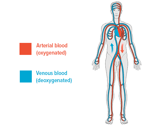
Veins are vessels that transport blood back to the heart. They’re part of the low pressure component of the cardiovascular system, as the pressure in our veins is significantly lower than in our arteries. Veins transport deoxygenated blood throughout the cardiovascular system, while arteries carry oxygenated blood as part of our pulmonary circulatory system. 

The veins in an adult’s body transport around 7,000 litres of blood back to the heart every day. The skeletal muscles are mainly responsible for pumping the blood through the veins, while the venous valves also play an important role in this process.

Most of the veins in the body run parallel to their arterial counterpart, and are therefore known as accompanying veins.

What’s the difference between veins and arteries? 

The entire human body is crisscrossed by two types of blood vessels: arteries and veins

Arteries transport oxygenated blood to organs, muscles, tissue and other areas where it is required. Arterial blood is lighter in colour than deoxygenated venous blood.

Most small and medium-sized veins have venous valves that prevent gravity from causing the blood to flow backwards. Arteries don’t have valves.

The human cardiovascular system

How does blood flow in a vein?

Blood is transported away from the heart when the heart beats. However, this pulse isn’t enough to carry the blood – once it has passed through the capillaries (tiny blood vessels)  – through the veins as well. The heart is essentially a pressure pump rather than a suction pump. It only plays a minor part in the return leg of the cycle.

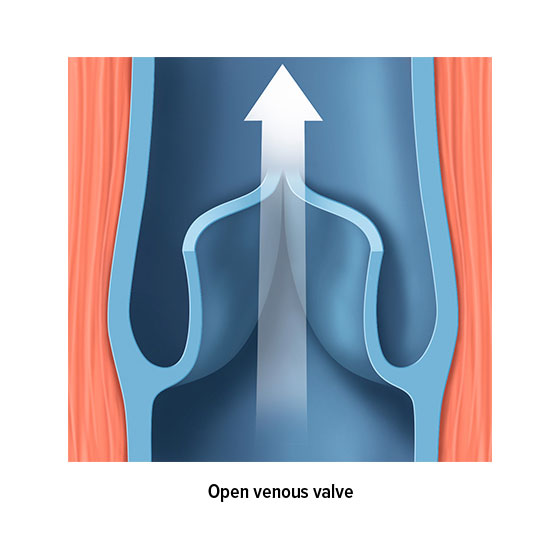
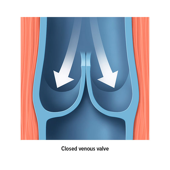
The venous valves are anatomically responsible for ensuring the blood flow. Most veins are divided into sections by valves. For example, there are up to 20 venous valves in the veins in our extremities, all of which work to ensure that our blood flows in the right direction – i.e. back to the heart. These valves function as a type of non-return valve. When pressure is applied to a section of vein, the valves open towards the heart and allow blood to pass along into the next section. However, the valves only open in one direction. This means that a healthy valve will prevent blood from flowing backwards.  

But how is pressure applied to the veins? Several mechanisms are responsible for ensuring that the blood flows back to the heart, and these are known collectively as venous pumps or skeletal-muscle pumps. When a muscle contracts, the vein closest to the muscle is also compressed, which allows the blood to continue on its journey back to the heart. The vessel wall acts as a counterpressure to the blood, and prevents the pressure in the vein from rising any further. When the muscle relaxes, the vein expands again. The resulting suction ensures that blood flows back from the deeper-lying sections of the vein and from the superficial veins closer to the skin into their deep-lying counterparts. Accompanying veins are compressed in a similar way, as the pulse passes through a nearby artery. Blood flow in the veins close to the heart is influenced by breathing, whereas blood flow in the abdomen is influenced by muscle contractions in the digestive tract.

This interplay of compression and venous valves ensures that blood can be transported around the body despite gravity – and this happens 24 hours a day, throughout our lives. Small wonder, then, that the valves and walls of the veins often stop functioning correctly as you grow older, and that venous disorders arise

The veins in the legs

The veins in the legs perform the biggest job in the circulatory system. They pump the blood from the lowest point in the body back to the heart without any breaks, and against gravity during the day. This is the price we pay for evolving to walk on two legs.

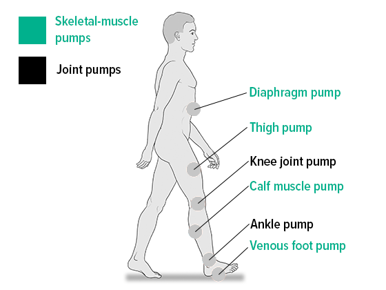
The “skeletal-muscle pump” of the leg muscles, also referred to as the “calf muscle pump”, performs the most important function when it comes to returning the blood to the heart. When the legs move, the muscles in the lower leg contract, acting like natural pumps that transport venous blood from the legs back up to the heart.

Even the smallest changes to a vein – for example, when it becomes dilated – can have a negative impact on the functioning of the venous valves and stop them from closing properly. This can cause the venous blood in the veins in the legs to accumulate and only flow slowly. This pooling of blood is initially perceived as “heavy” or “swollen” legs. The important task of returning blood to the heart can no longer be performed correctly, which, if untreated, can result in serious venous disease.

Here, we distinguish between the heavily branched superficial leg veins that run through the fatty subcutaneous tissue, the large, deep leg veins and the perforator veins that connect them.

You can find an overview of the various skeletal-muscle and joint pumps here:


Skeletal-muscle and joint pumps

Structure of the veins and venous system

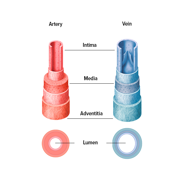
The thickest veins are the vena cava in the heart, which have a diameter of about 2 cm, while the thinnest venules (tiny venous vessels) are only about 15 μm in diameter. As is typical of all blood vessels, the venous wall is divided into three layers: 

Structure of the veins and arteries

1. Tunica interna (Intima)

This inner layer consists of a simple squamous epithelium – in other words, a single layer of flat mucosal cells sitting atop a basal membrane. This layer forms the venous valves.

2. Tunica media (Media)

This middle layer has a two-layer structure, with an inner spiral layer and a flatter outer layer. The smooth muscle cells are forced apart into strands by collagenous connective tissue, which makes the tunica media looser than in an artery.  

3. Tunica externa (Tunica adventitia)

This outer layer is a layer of connective tissue that fixes the vein in place. It houses nerves and – in the case of very large veins – smaller blood vessels that feed the larger vessels.

There are a few exceptions to this general venous anatomy: Certain large veins, such as the inferior vena cava, only have longitudinal muscle tissue, while the larger veins in the leg, such as the great saphenous vein (vena saphena magna) have a high hydrostatic pressure. The walls in these veins are similar in strength to those of arteries.

A venous valve is a protrusion on the wall of a vein. The valve works like a small sail. There are no venous valves in the veins in the head, intestines, spinal canal or the large veins close to the heart. In the arteries, the pumping pressure exerted by the heart is strong enough to prevent the blood from flowing backwards.

Open venous valve
Closed venous valve

Venous disorders: Symptoms

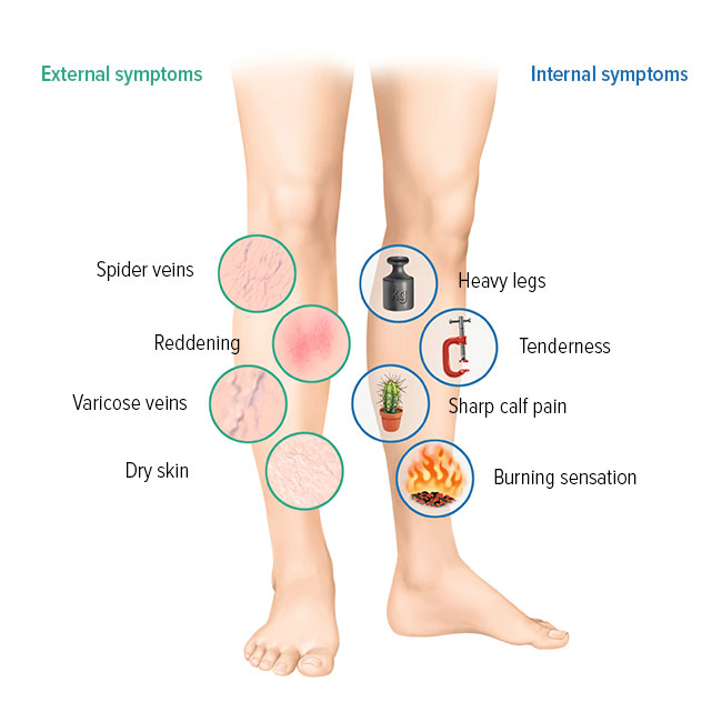
Nearly everyone has experienced that sensation: it’s the evening, and your legs feel as if they’re made of lead. It could be that you’ve had a tiring day with too much sitting or standing, but it could also be an indicator that you’re experiencing a venous disorder. Many symptoms are associated with venous disorders. If you want to find out what’s causing bulging veins, it’s also a good idea to look into the many other symptoms of venous disorders.


  • Highly visible veins on the foot
  • Thick veins in the legs
  • Bulging veins
  • Visible varicose veins and spider veins
  • Leg pain
  • Tired, heavy legs
  • Leg cramps at night
  • Itching
  • Burning
  • Tingling
  • Feeling of tightness
  • Dry, thin skin over the affected vein
  • Sharp pains
  • Pressure pain
  • Swelling in the ankles and calves
  • Reddening or other discolourations
  • Skin ulcerations in the ankle area
Symptoms of venous disorders

No symptoms – even mild ones – should be downplayed, especially if they recur. Without treatment, they can become worse and can even open the door to other serious venous diseases. Such symptoms should always be checked by a medical expert, preferably a specialist in the field of phlebology or dermatology.

Risk factors that contribute to venous disorders

There are a number of influencing factors that open the door to venous diseases. Here, we make a distinction between factors than we can influence and those that we can’t influence:

Various conditions

Just like on the road, queues can form in the veins. All it takes is one bottleneck to bring traffic to a standstill in the area. In the body, these bottlenecks can result from a small change to the vein that prevents the venous valve from fully closing, thereby reducing the blood flow. This valve deficiency is often the culprit for many venous disorders, but can also result from a disease affecting the circulatory system.

Spider veins

Lifestyle image spider veins

Spider veins

Spider veins are small, visible clusters of veins that form a web or fan shape in the epidermis (your outermost layer of skin). They are usually harmless and are more of a cosmetic issue, but can also indicate the onset of venous insufficiency.

Varicose veins (varicosis)

Lifestyle image varicose veins

Varicose veins (varicosis)

Varicose veins (varicosis) occur when the valves in a vein function inadequately and reduce the flow of blood back to the heart. Gravity causes blood to collect in the legs. This leads the venous walls to weaken, and the dilated vein becomes tortuous and knotty.

Venous insufficiency

Lifestyle image venous insufficiency

Venous insufficiency

Venous insufficiency refers to a disturbance to normal vein function, which usually occurs when the blood flow is impeded. Venous insufficiency normally affects the veins in the legs.

Inflammation of the vein (phlebitis)

Lifestyle image inflammation of the vein

Inflammation of the vein (phlebitis)

Phlebitis occurs when a venous blood vessel becomes inflamed. This condition can affect both superficial and deep veins. Both types of phlebitis are usually accompanied by the formation of blood clots.

Thrombosis


Lifestyle image thrombosis

Thrombosis

Thromboses occur as a result of a blood clotting disorder. When the blood suddenly clots in the vascular system, a blood clot (thrombus) can form on the vessel wall – this usually occurs in the veins, particularly in the deep veins in the legs. These deposits constrict the vessels or can even block them completely, which means that the blood can no longer flow efficiently through the veins to the heart. This is referred to as a deep vein thrombosis.

Venous leg ulcers


Lifestyle image venous leg ulcers

Venous leg ulcers

Venous leg ulcers are open, usually weeping wounds on the lower leg or foot that heal poorly, or sometimes don’t heal at all. They often develop as a result of a long-standing and untreated weakness of the veins.

Diagnosing venous disorders

Ultrasound scanning is the preferred method for imaging deep veins and accurately diagnosing diseased blood vessels. This also makes it possible to identify the speed and direction of the blood flow. Specialists can even examine the shape and structure of the veins with the help of duplex sonography, a pain-free procedure that comes with no side-effects.

In severe cases, additional scanning technologies such as magnetic resonance imaging (MRI) or computed tomography (CT scanning) may also be employed.


Preventing venous disorders

One of the best ways to prevent vein disease is to exercise – whether you enjoy it or not. Avoid extended, uninterrupted periods of standing or sitting. Variety is also essential. You don’t need to be an elite athlete: cycling, swimming or walking will absolutely do the trick. Taking cold showers and regularly raising your legs will also help. You should also minimise all risk factors that you can influence:

Maintain a balanced diet to avoid becoming overweight. Avoid heat sources such as saunas or direct sunlight. Give up alcohol and nicotine. Stick to wearing flat, comfortable shoes and avoid tight-fitting clothing. Explore alternatives to the contraceptive pill.

Vein exercises at a glance  – a brief overview 
 

Short vein exercises stabilise the vessel walls and promote circulation. Are you looking for exercises to do at home, at work or when you’re out and about? 

With Juzo’s vein exercises, it’s really easy to get your vein pumps going and thus support your health.

In the videos, you will find a selection of everyday exercises that are easy to perform. Repeat each exercise ten times.


Dealing with venous insufficiency

Once venous insufficiency sets in, preventive measures are often no longer sufficient to prevent the condition from deteriorating. Compression therapy is the core component in treating venous insufficiency. It helps to allow the valves in the veins (also known as venous valves) to fully close again, and thereby to transport the blood back to the heart.

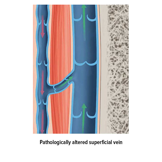
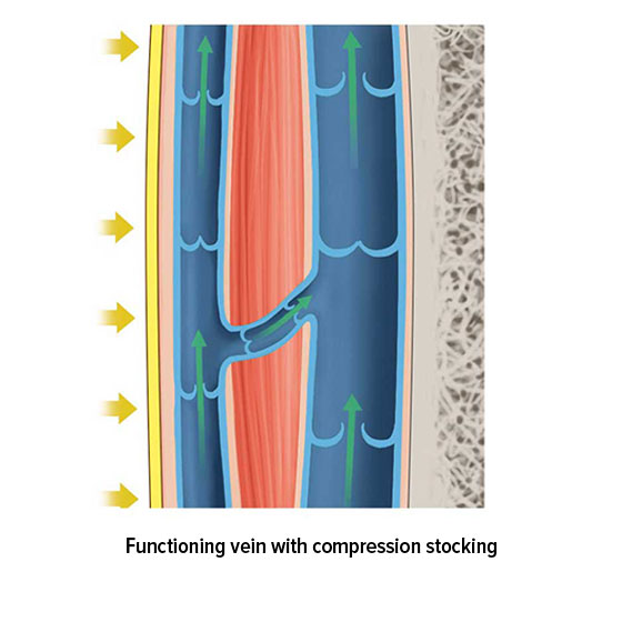
But how does compression therapy help with venous disorders? The core principle of compression therapy is to apply controlled external pressure to the tissue and the network of veins within it. This reduces the diameter of the veins, ensuring that valves that can’t currently fully close can do so again, and thereby function as non-return valves. The pressure applied by the compression stockings is lower closer to the heart, which also helps with transporting the blood in that direction. Compression stockings work particularly well in combination with exercise, which allows the calf muscle pump to function as effectively as possible.

Compression therapy can be supplemented with medication, as well as various other treatments or surgical procedures.

Pathologically altered superficial vein
Functioning vein with compression stocking

Products for treating venous disorders

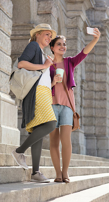
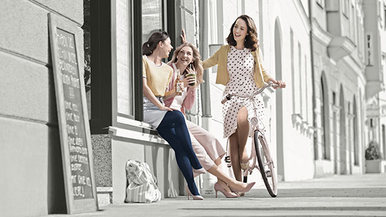
Three women in the city. They all wear compression stockings

Juzo Inspiration

Lifestyle combined with an elegant design.
 

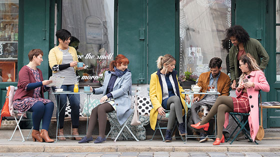
Friends in a café. Some of them are wearing compression stockings.

Juzo Soft

Super soft for feel good moments.
 

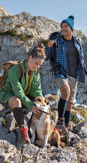
Three men with a dog in the mountains. The men are wearing compression stockings on their lower legs.

Juzo Adventure

The all-rounder for active men.
 

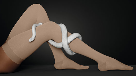
One leg with a compression stocking. A snake winds around the leg.

Juzo Move

Patented knit for easy donning and doffing.
 

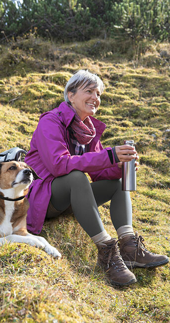
A woman sits on a meadow. She is wearing Juzo Dynamic compression tights. A dog is sitting next to her.

Juzo Dynamic

Reliably durable.
 

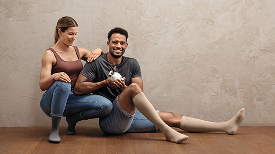
A man and a woman are sitting on the floor. Both are wearing compression stockings on their lower legs. The man is holding a small bunny in his hands

Juzo Intenso

The strong stocking with a soft side
 

A product image of Juzo Basic compression tights

Juzo Basic

Proven quality for everyday life.
 

A product image of a Juzo Ulcer Pro

Juzo Ulcer Pro

The compression stocking system for the treatment of venous leg ulcers